31 August 2026, Volume 46 Issue 8
    

  • Select all
    |
    Review & Monography
  • HU Xiao-wen, YUAN Song, LIU Bo, LI Xiu-ji, WANG Song-yuan, LIU Yang, SUN Hui-min, CHEN Hua
    Chinese Journal of Pharmaceutical Analysis. 2026, 46(8): 1373-1379. https://doi.org/10.16155/j.0254-1793.2025-0508
    Abstract ( )   Knowledge map   Save
    Objective: To develop a high-precision and interpretable drug mutagenicity prediction model based on AI to assist in new drug development and pharmaceutical regulatory oversight. Methods: Mutagenicity data from various sources were collected and integrated to establish a standardized dataset of small organic molecules. A graph neural network framework was employed to train the model on the training set, while an external test set was used to evaluate its generalization capability. The applicability domain of the model was defined based on molecular fingerprint features and the convex hull algorithm was employed to assess prediction reliability. Additionally, an atomic perturbation-based analysis was conducted to investigate the prediction mechanisms of the model. Finally, a web server was developed as an internal mutagenicity prediction platform. Results: The optimized AI model achieved the accuracy of 0.828, recall of 0.863, precision of 0.848, and an area under the receiver operating characteristic curve of 0.907 on the training set. On the external test set, these metrics were 0.751, 0.789, 0.798, and 0.775, respectively. Furthermore, the model successfully identified key toxic structures or groups in compounds, such as sulfonate esters, nitrosamines, halogenated alkanes, and hydrazines, and effectively distinguished between samples within and outside the applicability domain. Furthermore, an online prediction platform was established, enabling toxicity assessment via molecular structures. Conclusion: The proposed model not only accurately predicts compound mutagenicity but also visualizes the potential risk sites, demonstrating robust generalization ability and practical utility. This study provides an efficient AI-driven solution for the early screening of genotoxic impurities.
  • YAN Jian-gong, FANG Wen-liang, GUO Ri-xin, ZHOU Ya-nan, LIU Jing-jing, LIU Jing, WEI Feng
    Chinese Journal of Pharmaceutical Analysis. 2026, 46(8): 1380-1389. https://doi.org/10.16155/j.0254-1793.2025-0374
    Abstract ( )   Knowledge map   Save
    Caffeoylquinic acids are a class of natural components widely distributed in medicinal plants in nature, exhibiting multiple biological activities. The activities of caffeoylquinic acids are closely related to their structures. The diversity of hydroxyl esterification sites and spatial conformations in their structures makes it difficult to accurately distinguish isomers by a single conventional identification approach. Therefore, the combined application of multiple identification approaches is usually required to ensure accurate structural determination. Although research reports on caffeoylquinic acids are increasing, a comprehensive summary of their structural analysis and biological activities is still lacking. Based on the research reports on caffeoylquinic acids over the past two decades, this paper summarized the results on structural analysis, biological activities and quality control markers, aiming to provide a reference for the application of caffeoylquinic acid isomers in the quality control of traditional Chinese medicine.
  • Ingredient Analysis
  • AILI Re-ze-ye, HONG Yu-wen, GU Yue, TAO Chu-jun, QIU Feng
    Chinese Journal of Pharmaceutical Analysis. 2026, 46(8): 1390-1403. https://doi.org/10.16155/j.0254-1793.2025-0665
    Abstract ( )   Knowledge map   Save
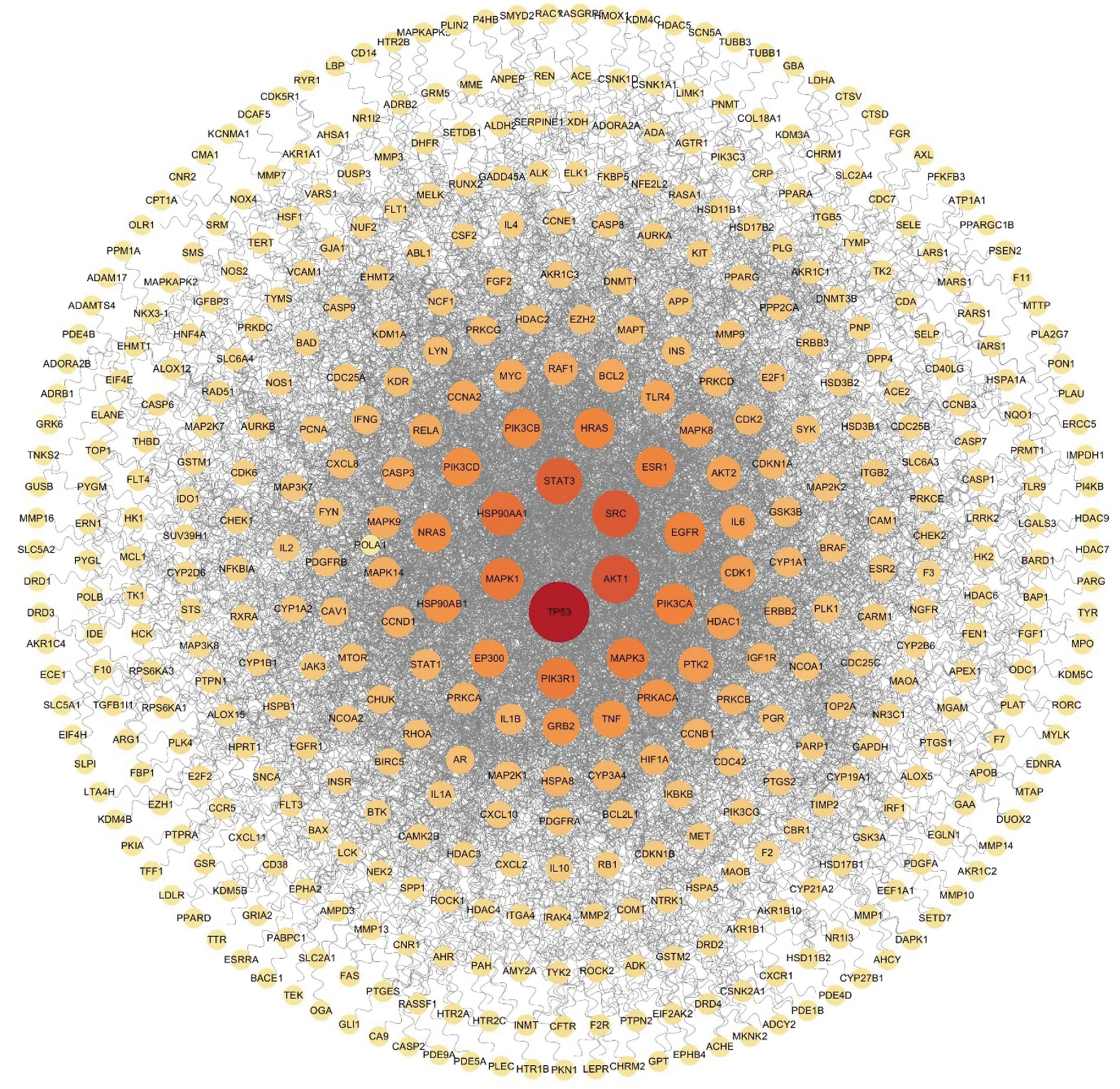
    Objective: To predict the potential quality markers (Q-markers) of Tongluo Mingmu capsules based on network pharmacology and establish an ultra-high performance liquid chromatography-ultraviolet detection (UPLC-UV) method for their simultaneous quantification, thus improving the quality standard of Tongluo Mingmu capsules. Methods: The “component-target-metabolic pathway” interaction data of Tongluo Mingmu capsules were collected through network pharmacology approaches, and the resulting network was constructed for visualization. UPLC-UV was performed through an Acquity UPLC HSS T3 column (100 mm×2.1 mm, 1.8 μm) with a column temperature of 30 ℃. The mobile phase consisted of 0.1% formic acid aqueous solution (A)-acetonitrile (B) for gradient elution with the flow rate of 0.30 mL · min-1 at the detection wavelength of 261 nm. The injection volume was set to be 5 μL. The UPLC-UV method was then used to simultaneously quantify 14 components (puerarin, paeoniflorin, luteolin, formononetin, aloeemodin, physcion, rhein, emodin, chrysophanol, ecliptasaponin A, specnuezhenide, ginsenoside Rg1, daidzein, and wedelolactone) in Tongluo Mingmu capsules. Results: A total of 35 potential Q-markers of Tongluo Mingmu capsules were screened out, which could bind to protein targets such as TP53 to regulate signaling pathways including the PI3K-Akt signaling pathway. All method validation indexes of the established UPLC-UV method met the methodological requirements. In three batches of Tongluo Mingmu capsule samples, the content ranges of puerarin, paeoniflorin, luteolin, formononetin, aloe-emodin, physcion, rhein, emodin, chrysophanol, ecliptasaponin A, specnuezhenide, ginsenoside Rg1, daidzein, and wedelolactone were 0.350-0.376 mg per capsule, 2.198-2.666 mg per capsule, 0.187-0.249 mg per capsule, 0.040-0.046 mg per capsule, 0.078-0.086 mg per capsule, 0.214-0.272 mg per capsule, 0.142-0.152 mg per capsule, 0.148-0.162 mg per capsule, 0.288-0.321 mg per capsule, 0.409-0.491 mg per capsule, 2.630-3.220 mg per capsule, 0.021-0.028 mg per capsule, 0.019-0.026 mg per capsule, and 2.108-2.566 mg per capsule, respectively. Conclusion: Network pharmacology provides a theoretical basis for the screening of Q-markers in Tongluo Mingmu capsules. The established UPLC-UV method can be used to improve the quality standard of Tongluo Mingmu capsules.

  • LÜ Feng-jiao, LIU Min, WEN Yun, XIE Xiao-lan
    Chinese Journal of Pharmaceutical Analysis. 2026, 46(8): 1404-1411. https://doi.org/10.16155/j.0254-1793.2025-0361
    Abstract ( )   Knowledge map   Save
    Objective: To establish a high-performance liquid chromatography (HPLC) method for the determination of diosmetin in thermosensitive liposomes and investigate its in vitro release behavior. Methods: HPLC was conducted with a Supersil ODS2 column (250 mm×4.6 mm, 5 μm), the detection wavelength of 343 nm, and the injection volume of 20 μL. Single-factor experiments were carried out to investigate the influences of mobile phase methanol-water ratio by volume, flow rate, and column temperature and optimize the chromatographic conditions. With 1% Tween 80 aqueous solution as the release medium, the in vitro release behavior of diosmetin-loaded thermosensitive liposomes at different temperatures was investigated via dialysis coupled with constant-temperature shaking. The cumulative release rate was calculated, and Origin software was adopted to fit drug release models. Results: The optimized chromatographic conditions were as follows: a mobile phase composed of methanol and water at a volume ratio of 75 ∶ 25, a flow rate of 1 mL · min-1, and a column temperature of 35 ℃. Diosmetin exhibited a good linear relationship between peak area and concentration within the range of 0.1-50 μg · mL-1, and the regression equation was A=86.489C-2.142 2 (r=1.000). The limit of detection was 0.6 ng, and the limit of quantitation was 1.8 ng. The spiked recoveries at high, medium, and low concentrations were all above 95%, and the RSD values were all less than 2%. In vitro release experiments revealed that diosmetin-loaded thermosensitive liposomes exhibited obvious temperature responsiveness. Compared with free diosmetin, the liposomes showed slow drug release at 37 ℃, whereas the release rate rose significantly at 42 ℃ and 53 ℃. The release behavior at 37, 42, and 53 ℃ all complied with the first-order kinetic model. Conclusion: This method is simple, stable, and efficient, and is thus suitable for the in vitro release evaluation of diosmetin-loaded thermosensitive liposomes. Furthermore, the prepared liposomes exhibit heat-triggered drug release behavior and sustained-release properties, and their release behavior is primarily dominated by diffusion.
  • WU Yue, MA Chi, KONG Ling-jie, ZHANG Wei-dong
    Chinese Journal of Pharmaceutical Analysis. 2026, 46(8): 1412-1419. https://doi.org/10.16155/j.0254-1793.2025-0785
    Abstract ( )   Knowledge map   Save
    Objective: To establish an ultra-performance liquid chromatography-triple quadrupole mass spectrometry (UPLC-QQQ MS/MS) method for the simultaneous determination of nine blood-absorbed components in Qijiao Shengbai capsules, including 7-hydroxy-4-benzopyrone, calycosin, formononetin, medicarpin, p-coumaric acid, daidzein, maackiain, emodin, and icaritin. Methods: Chromatographic separation was performed on a Waters Acquity UPLC HSS T3 column (150 mm×2.1 mm, 1.8 μm) using gradient elution, with 0.1% formic acid in water (A) and acetonitrile (B) as the mobile phase and column temperature at 30 ℃. The flow rate was 0.2 mL · min-1,and the injection volume was 2 μL. Mass spectrometry was carried out using an electrospray ionization (ESI) source, and both positive- and negative-ion scanning were performed in multiple reaction monitoring (MRM) mode. Results: The established method showed good linear relationships (r≥0.998 1) and demonstrated good precision, repeatability, and stability (RSD<5.0%). The average recoveries (n=6) of the nine blood-absorbed components ranged from 97.3% to 107.2% with RSDs under 5.0%. The content ranges of the nine compounds in three batches of Qijiao Shengbai capsules were 0.271 2-0.275 3 μg · g-1, 139.9-146.5 μg · g-1, 42.31-43.80 μg · g-1, 5.817-5.946 μg · g-1, 6.740-7.145 μg · g-1, 1.206-1.246 μg · g-1, 42.83-44.78 μg · g-1, 0.364 4-0.387 0 μg · g-1, and 2.787-2.967 μg · g-1. Conclusion: The established method is specific, sensitive, and stable. It can be used to simultaneously determine the contents of nine blood-absorbed components in Qijiao Shengbai capsules, providing a scientific basis for its quality control and safe medication.
  • HUANG Yan-chang, HUANG Yu-tong, XUE He-fei, XING Yan-ping, YANG Yan-yun, KANG Ting-guo, DOU De-qiang, XU Liang
    Chinese Journal of Pharmaceutical Analysis. 2026, 46(8): 1420-1430. https://doi.org/10.16155/j.0254-1793.2025-0462
    Abstract ( )   Knowledge map   Save
    Objective: To investigate the histological structural differences and the spatial distribution characteristics of saponins in the roots and rhizomes of Pulsatilla chinensis (Bunge) Regel, Pulsatilla cernua (Thunb.) Bercht. & J. Presl, and Pulsatilla turczaninovii Krylov & Serg. by combining microscopy and matrix-assisted laser desorption/ionization mass spectrometry imaging (MALDI-MSI). Methods: The histological structural characteristics of the roots and rhizomes of three Pulsatilla species were observed and compared by microscopy. An OLYMPUS microscopic imaging system and a BX-51 biological microscope equipped with a DP-72 microscope digital camera and polarizing filters were used for observation, and the microscopic features were documented. For MALDI-MSI analysis, tissue sections were prepared through a Leica CM1950 cryostat, and DHB matrix was applied with a TM-Sprayer matrix sprayer. The matrix-coated ITO slides were placed on the mass spectrometer target plate, and tissue regions of interest were selected by DataImaging. Under identical laser energy, the TimsTOF fleX mass spectrometry system was used to identify and detect tissue samples, and the in situ spatial distribution of 21 saponins, including the marker component pulchinenoside B4 specified in the Pharmacopoeia of the People’s Republic of China, was visualized across different tissue regions of the roots and rhizomes. Results: Microscopic observations revealed that the roots of all the three species possessed a distinct endodermis (with prominent casparian strips), while the rhizomes contained a pith or fissures. Significant interspecies differences were observed in the number of xylem bundles and the distribution of phloem fibers. MALDI-MSI results revealed that among the 21 saponins, the majority (17) of saponins were predominantly enriched in the cortical tissue, while 4 were undetected. Some saponins (pulchinenoside D and pulchinenoside E2) exhibited a unique ring-shaped distribution pattern in the endodermal region of the roots or rhizomes, most prominently in the roots of P. chinensis and P. cernua. Pulchinenoside E showed higher distribution in the xylem. Conclusion: This study reveals the specific spatial distribution patterns of saponins (cortex enrichment and endodermal ring distribution) in the roots and rhizomes of these three Pulsatilla species and clarifies the interspecies differences in their microscopic tissue structures. This study reveals histochemical differences among three Pulsatilla species and between their roots and rhizomes, providing a histochemical basis for species identification and the scientific evaluation and development of medicinal resources of this genus.
  • JIA Zhi-xin, MA Tian-xin, ZHANG Ping, LIU Meng-jiao, KANG Rong, YANG Jian-bo, LIU Wei, YU Kun-zi, CHEN Jia, CHENG Xian-long, WEI Feng
    Chinese Journal of Pharmaceutical Analysis. 2026, 46(8): 1431-1438. https://doi.org/10.16155/j.0254-1793.2025-0491
    Abstract ( )   Knowledge map   Save
    Objective: To screen the differential components between the root and vine of Pueraria lobata (Willd.) Ohwi by high-resolution mass spectrometry combined with omics methods, isolate one marker component, and develop a content determination method for this component, thereby furnishing scientific data for the quality evaluation of P. lobata. Methods: The mass spectrometry (MS) data were collected through a Q Exactive high-resolution MS instrument. The MS examination was carried out with ESI source in the negative ion mode, a spray voltage of 3.5 kV, a capillary temperature of 350 ℃, a sheath gas flow rate of 35 arb, an auxiliary gas flow rate of 10 arb, an acquisition range of m/z 150-1 500, and a resolution of 70 000. MS/MS data were acquired under dd-MS2/dd-SIM mode at a resolution of 17 500. Differential components were screened via omics combined with multivariate statistical analysis. One of the differential components was isolated, and its structure was confirmed by MS and NMR data. Gradient elution was performed through an Agilent SB-C18 chromatographic column (250 mm×4.6 mm, 5 μm) at a column temperature of 30 ℃, with the mobile phase being acetonitrile-0.1% formic acid water at a flow rate of 1.0 mL · min-1. The detection wavelength was 254 nm, and the injection volume was 5 μL. An HPLC method for the marker compound was established and validated, and the established method was employed to determine the content of this differential component in P. lobata. Results: This differential component exhibited good linearity between peak area and concentration within the range of 0.97-19.40 μg · mL-1, with a correlation coefficient (r) of 0.999 7. The relative standard deviations (RSDs) were 1.4%, 2.1%, and 2.5% for method precision, repeatability, and stability, respectively. The average spiked recovery rate was 92.2%, with the RSD of 2.5%. The content of this differential component in the root and vine of P. lobata varied within the ranges of 0.033 9% to 0.066 4% and ND to 0.009 5%, respectively. There was a significant difference in the component content between root and vine (P<0.000 1 and a fold change of 12). Conclusion: This study employs high-resolution MS and omics to identify the differential components between the root and vine of P. lobata. Subsequently, a specific compound is isolated, and a robust content determination method is developed. The methodological validation results demonstrate excellent precision, high repeatability, and superior separation efficiency. The findings of this study offer valuable data insights to support the quality evaluation of P. lobata.
  • LIU Xian-lei, MENG Wei-ting, LI Jin-yan, WANG Xiao-yan, ZHANG Hui, ZHANG Ming-liang, CHEN Xiao-fei, SUN Wen-jing, LI Wei-xia, TANG Jin-fa
    Chinese Journal of Pharmaceutical Analysis. 2026, 46(8): 1439-1448. https://doi.org/10.16155/j.0254-1793.2025-0538
    Abstract ( )   Knowledge map   Save
    Objective: To establish the quality evaluation method of Gancao Ganjiang decoction (Glycyrrhizae Radix et Rhizoma Praeparata Cum Melle-Zingiberis Rhizoma; Glycyrrhizae Radix et Rhizoma Praeparata Cum Melle-Zingiberis Rhizoma Praeparatum) formula granules, and to comprehensively evaluate the quality uniformity and stability of the formula granules from different manufacturers. Methods: The chromatographic column Acquity UPLC® BEH C18 at 30 ℃ was used for gradient elution with the mobile phase composed of acetonitrile and 0.05% phosphoric acid at a flow rate of 0.30 mL · min-1. The detection wavelength was 235 nm. Results: The linear relationship was good (0.950-1.000) for each component. The average recovery rates were 97.9%-100.6%, and the relative standard deviations (RSDs) were 2.6%-5.0%. The content of components in 28 batches of samples from three manufacturers was determined. The statistical analysis of chemometrics showed that 6-gingerol, liquiritigenin, and 6-shogaol contributed the most to the content difference of Gancao Ganjiang decoction (Glycyrrhizae Radix et Rhizoma Praeparata Cum Melle-Zingiberis Rhizoma) formula granules among manufacturers, and 6-gingerol, glycyrrhizic acid, liquiritigenin, 6-shogaol, and zingerone contributed the most to the content difference of Gancao Ganjiang decoction (Glycyrrhizae Radix et Rhizoma Praeparata Cum Melle-Zingiberis Rhizoma Praeparatum) formula granules among the three manufacturers. The Gancao Ganjiang decoction (Glycyrrhizae Radix et Rhizoma Praeparata Cum Melle-Zingiberis Rhizoma) formula granules and Gancao Ganjiang decoction (Glycyrrhizae Radix et Rhizoma Praeparata Cum Melle-Zingiberis Rhizoma Praeparatum) formula granules from the same manufacturer were grouped into one group, respectively. Glycyrrhizic acid and 6-gingerol contributed the most to the content difference between the two formula granules. Conclusion: A UPLC method was established for the simultaneous determination of 8 components (isoliquiritin apioside, glycyrrhizic acid, liquiritigenin, zingerone, 6-shogaol, 10-gingerol, 8-gingerol, and 6-gingerol) in Gancao Ganjiang decoction formula granules. This method is fast and stable, and provides a scientific basis for the overall quality evaluation of the formula granules.
  • Metabolism Analysis
  • GAN Zhen, LI Jin-fa, WANG Yu, CHEN Si-yuan, CHANG Jun-min
    Chinese Journal of Pharmaceutical Analysis. 2026, 46(8): 1449-1458. https://doi.org/10.16155/j.0254-1793.2026-0069
    Abstract ( )   Knowledge map   Save
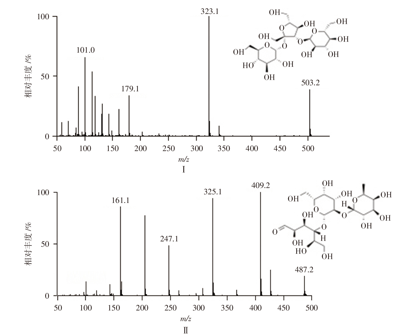
    Objective: To establish and validate an ultra-performance liquid chromatography-tandem mass spectrometry method, and to systematically investigate the pharmacokinetics, tissue distribution, and excretion of Melezitose in rats. Methods: Biological samples were prepared by protein precipitation with acetonitrile, and 2’-fucosyllactose was used as the internal standard. Quantitative analysis was performed by UPLC-MS/MS in the negative ESI mode with multiple reaction monitoring (MRM). Chromatographic separation was performed on a Waters Acquity UPLC BEH Amide column (2.1 mm×150 mm, 1.7 μm), using 0.1% ammonia aqueous solution (A) and acetonitrile (B) as the mobile phase with gradient elution (0-5 min, 85%B→75%B; 5-10 min, 75%B→65%B; 10-15 min, 65%B→70%B). The flow rate was 0.25 mL · min-1, the column temperature was 40 ℃, and the injection volume was 10 μL. Mass spectrometric detection was carried out on a Waters TQD triple quadrupole with an ESI source in negative ion mode, using MRM for quantification. The capillary voltage was 2.8 kV, the cone voltage was 30 V, the source temperature was 120 ℃, the desolvation temperature was 450 ℃, the nitrogen flow rate was 800 L · h-1, and argon was used as the collision gas. The method was validated for specificity, linearity, accuracy, precision, recovery, matrix effect, stability, and dilution integrity. Based on the validated method, three intragastric administration groups of melezitose (200 mg · kg-1, 400 mg · kg-1, 800 mg · kg-1) and intravenous administration group (200 mg · kg-1) were established to investigate its pharmacokinetics, tissue distribution, as well as urinary and fecal excretion characteristics in rats. Results: Melezitose exhibited good linearity over the concentration range of 0.1-20 μg · mL-1, with a lower limit of quantification of 0.1 μg · mL-1. All validation parameters met the requirements for quantitative analysis of biological samples. The absolute oral bioavailability of melezitose after intragastric administration ranged from 6.70% to 7.34%. The tissue distribution results demonstrated that the highest exposure level of melezitose occurred in the small intestine, followed by a rapid decline. The excretion study revealed that approximately 60% of intragastrically administered melezitose was excreted as the parent compound in feces within 36 h, while the urinary excretion accounted for less than 3% of the dose. Conclusion: The established UPLC-MS/MS method is sensitive, accurate, and robust, and is suitable for the systematic quantification of melezitose in rats.
  • Activity Analysis
  • CHEN Si-yu, XU Yang, CHEN Sheng-nan, ZHANG Ya-jie, BAO Hui-wei
    Chinese Journal of Pharmaceutical Analysis. 2026, 46(8): 1459-1471. https://doi.org/10.16155/j.0254-1793.2026-0022
    Abstract ( )   Knowledge map   Save
    Objective: This study aimed to establish a high-speed countercurrent chromatography (HSCCC) method for the separation of catechin (CAT) and epicatechin (EC) from Catechu, and to investigate the effects of CAT and EC on skin pruritus and vascular permeability in mice. Methods: High-purity CAT and EC were successfully isolated from Catechu by HSCCC with optimized separation parameters. An Agilent TC-C18 column (250 mm×4.6 mm, 5 μm) was adopted for high-performance liquid chromatography analysis. Methanol-water (30 ∶ 70) served as the mobile phase under isocratic elution conditions, with a flow rate of 1.0 mL · min-1, injection volume of 10 μL, column temperature of 30 ℃, and detection wavelength set at 280 nm. Pruritus mouse models induced by histamine (HA), chloroquine (CQ) and 2,4-dinitrofluorobenzene (DNFB) were established to evaluate the antipruritic efficacy of CAT and EC as well as their influences on cutaneous vascular permeability. Results: The purity of CAT and EC separated by HSCCC was 95.2% and 96.4%, with recovery rates of 83.4% and 84.1%, respectively. In mouse models of acute and chronic pruritus, the low-dose CAT group (CAT-L, 200 mg · kg-1), high-dose CAT group (CAT-H, 400 mg · kg-1), low-dose EC group (EC-L, 50 mg · kg-1), and high-dose EC group (EC-H, 100 mg · kg-1) all significantly reduced the frequency of scratching, decreased skin vascular permeability, and inhibited the release of inflammatory factors in a clear dose-dependent manner. The high-dose groups exhibited significantly better effects on antipruritus and improving vascular permeability than the low-dose groups. Conclusion: This study is the first to establish and optimize an HSCCC system for the separation of CAT and EC from Catechu, and the first to systematically evaluate their antipruritic activities and characteristics in acute and chronic pruritus models. The method is efficient, stable, simple, and yields high-purity products, making it suitable for the separation and preparation of CAT and EC from Catechu. Both CAT and EC show significant therapeutic effects on acute and chronic pruritus, providing new directions and experimental references for the research and development of natural drugs against skin pruritus.
  • Bioassay
  • SONG Ming-hui, WEI Ze-yu, GUAN Zi-qiu, FAN Yi-ling, SHAO Hong
    Chinese Journal of Pharmaceutical Analysis. 2026, 46(8): 1472-1482. https://doi.org/10.16155/j.0254-1793.2025-0456
    Abstract ( )   Knowledge map   Save
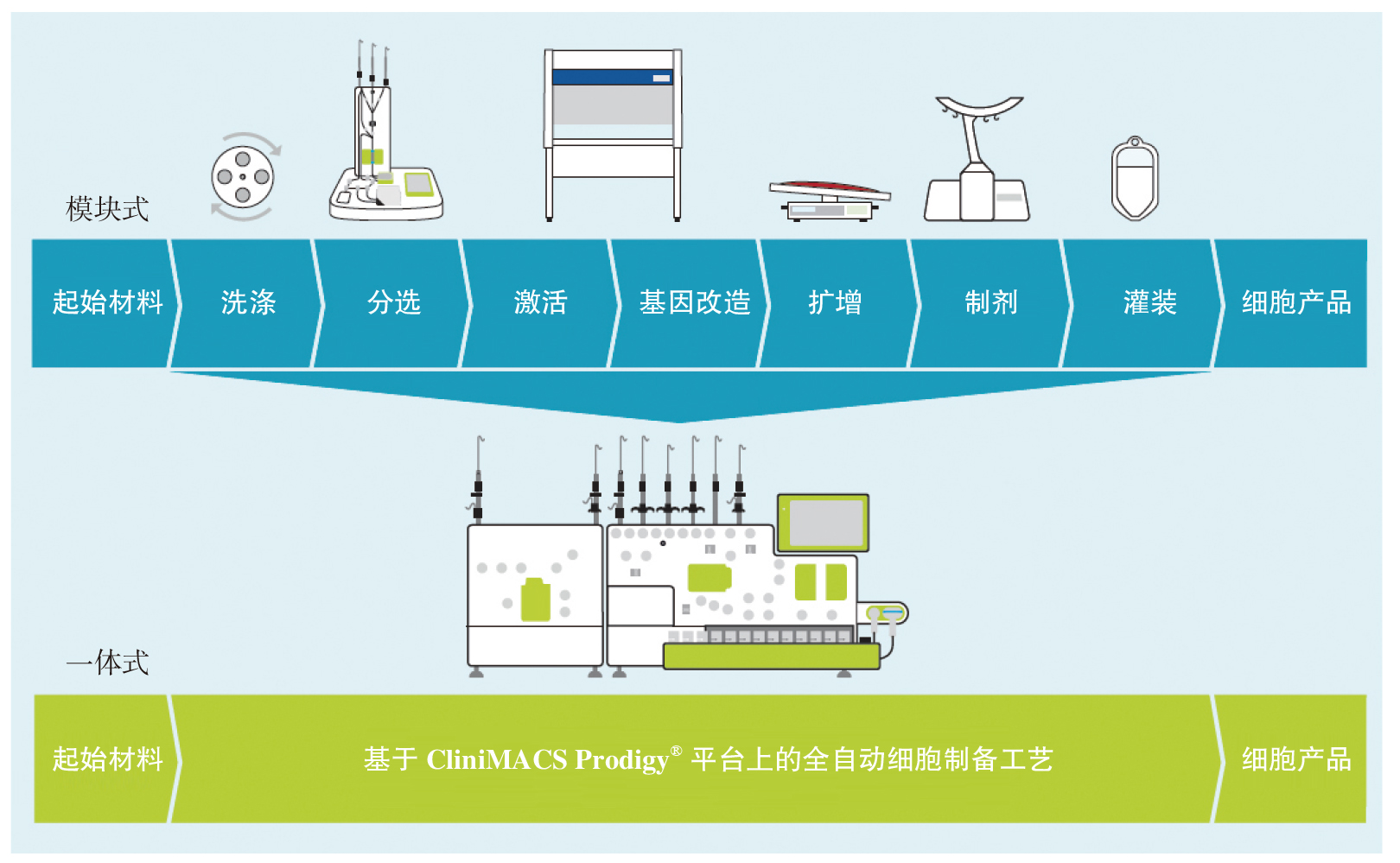
    Objective: To systematically investigate the design of aseptic process simulation intervention activities applicable to cell therapy medicinal products (CTMPs), represented by chimeric antigen receptor T-cell (CAR-T) products, identify potential contamination risks during the manufacturing process, and improve aseptic assurance capabilities. Methods: Regulations and technical guidelines issued by major regulatory agencies in China and abroad were systematically reviewed, and differences in their applicability to CTMPs were analyzed. Based on a scientific risk assessment approach, an intervention risk assessment model was applied to identify and classify intervention activities during the CAR-T manufacturing process. Key intervention points were then incorporated into the design of aseptic process simulation trials to develop a targeted simulation plan. Results: This study identified key intervention activities in the aseptic process simulation design of CTMPs represented by CAR-T products, established a quantifiable risk assessment matrix for intervention activities, and provided a scientific approach for identifying potential contamination risks during CTMPs manufacturing processes, thereby enhancing the effectiveness of aseptic process simulation trials for CTMPs. Conclusion: This study provides a technical basis for optimizing the aseptic assurance system of CTMPs represented by CAR-T products, offers guidance for the development of standardized practices in related industries, and provides a reference for the revision of relevant technical guidelines.
  • CUI Chun-bo, DU Jia-liang, LI Xin, NI Yong-bo, XU Gang-ling, YU Chuan-fei
    Chinese Journal of Pharmaceutical Analysis. 2026, 46(8): 1483-1489. https://doi.org/10.16155/j.0254-1793.2025-0493
    Abstract ( )   Knowledge map   Save
    Objective: To establish a method for determining the content of poloxamer 188 added in panitumumab based on high performance liquid chromatography-evaporative light scattering detector (HPLC-ELSD). Methods: The content of poloxamer 188 was determined through an Oasis wax column (20 mm×2.1 mm, 30 µm/80 Å) at a column temperature of 30 ℃. Gradient elution was performed with 0.2% formic acid aqueous solution as mobile phase A and acetonitrile containing 0.2% formic acid as mobile phase B at a flow rate of 1.0 mL · min-1. The ELSD was used. Results: The specificity of the method was strong. In the range of 5 to 80 μg · mL-1, the logarithm of the content of poloxamer 188 was linearly correlated with the logarithm of the peak area, with a coefficient of determination r2>0.99. The recovery rates of the six validation solutions with different poloxamer 188 content (5 μg · mL-1,10 μg · mL-1, 20 μg · mL-1, 40 μg · mL-1, 60 μg · mL-1, and 80 μg · mL-1) were in the range of 90.0%-110.0%. Three independent measurements were carried out on three different working days for each of the six validation solutions mentioned above, and the RSDs for their concentrations were 4.4%, 5.5%, 2.3%, 0.66%, 1.6%, and 1.6%, respectively. The injection was repeated six times for the validation solution at 40 μg · mL-1, and the RSD of their poloxamer 188 content was 0.60%. The RSD of the poloxamer 188 content was 0.80% for the validation solution at 40 μg · mL-1, which was assayed after storage for 0 h and 24 h at room temperature, respectively. Conclusion: An HPLC-ELSD method for content deter mination of poloxamer 188 in monoclonal antibody drugs was successfully developed, demonstrating good reproducibility, specificity, precision, and accuracy, which can provide a reliable technical basis for the quantitation and quality control of poloxamer 188 in monoclonal antibody drugs.
  • Safety Monitoring
  • LI Xiao-xin, CAI Jian-lei, LI Qiong-xia, LU Jun-qi
    Chinese Journal of Pharmaceutical Analysis. 2026, 46(8): 1490-1497. https://doi.org/10.16155/j.0254-1793.2025-0413
    Abstract ( )   Knowledge map   Save
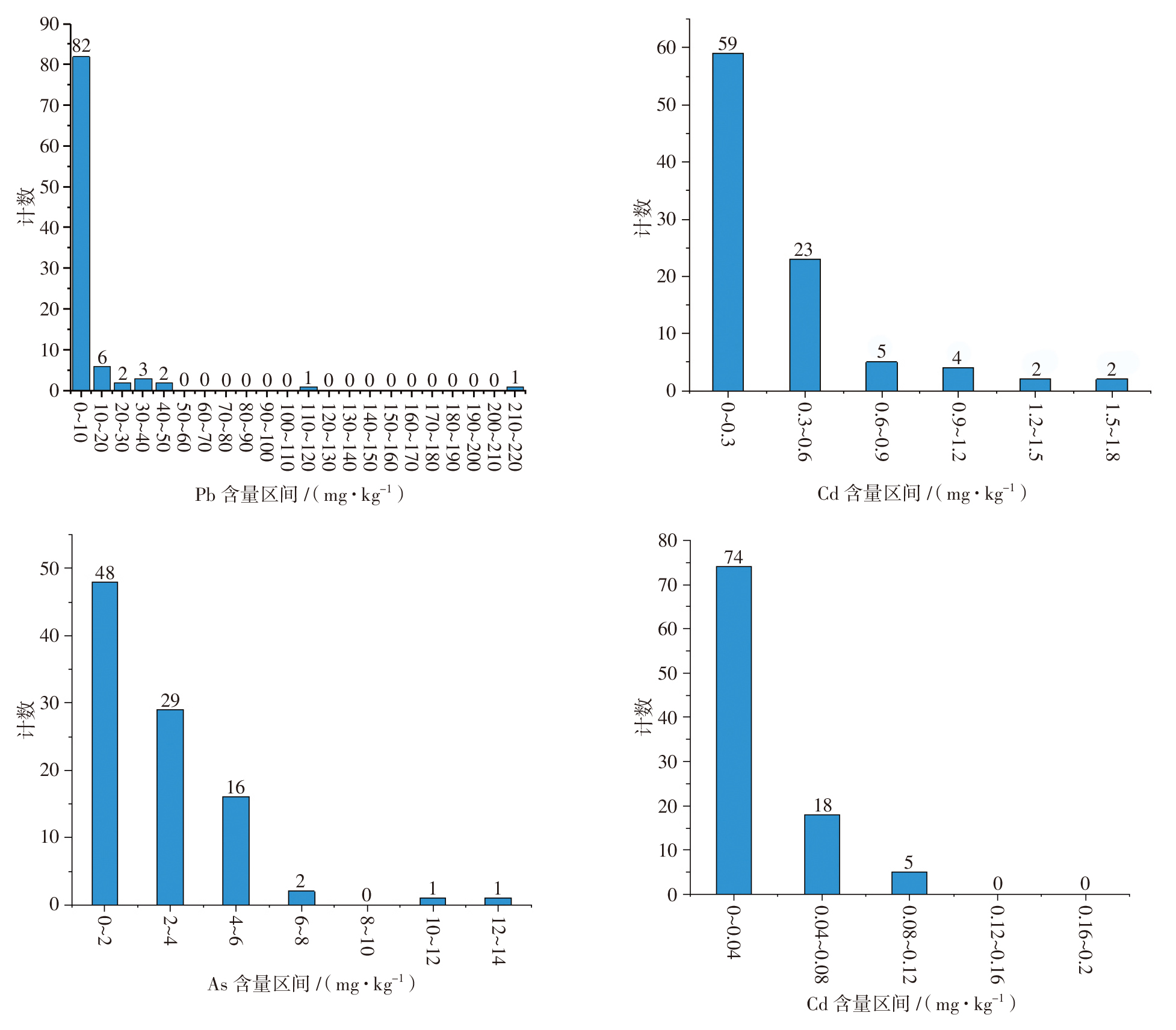
    Objective: To evaluate the residual levels and health risks of heavy metals and harmful elements in blood-activating traditional Chinese medicine (TCM) preparations containing insect-based drugs, thus providing a scientific basis for clinical safe medication and quality control of TCM preparations. Methods: Inductively coupled plasma mass spectrometry (ICP-MS) was employed to determine the content of lead (Pb), cadmium (Cd), arsenic (As), mercury (Hg), copper (Cu), vanadium (V), cobalt (Co), and nickel (Ni) in 97 batches of blood-activating TCM preparations containing insect-based drugs. Multivariate statistical analysis, one-way analysis of variance (ANOVA), principal component analysis (PCA), and health risk assessment models were combined to systematically evaluate the non-carcinogenic and carcinogenic risks of these elements. Results: The 75th percentile (P75) values of Pb, Cd, As, Hg, Cu, V, Co, and Ni in the 97 batches of TCM preparations were 5.04, 0.365, 3.80, 0.039 0, 30.2, 1.14, 1.38, and 4.63 mg · kg-1, respectively. Significant differences in the hazard levels of various elements were observed among different varieties. Multivariate statistical analysis, one-way ANOVA, and robustness testing all yielded P-values less than 0.05 and effect sizes greater than 0.04. PCA results indicated that Cd, As, Hg, and Cu were the major contributors to health hazards. The LCR values for carcinogenic risks in all varieties of TCM preparations were less than 1×10⁻⁶, within an acceptable range. Among non-carcinogenic risks, Pb in Tongxinluo capsules and Qizhi Tongluo capsules showed the hazard index (HI) greater than 1, which warranted attention. Conclusion: The carcinogenic risks of 97 batches of TCM preparations are relatively low. In terms of non-carcinogenic risks, the exposure to Pb in Tongxinluo capsules and Qizhi Tongluo capsules deserves further attention. This study provides a reference for the safety evaluation of TCM preparations.
  • ZHANG Lei, FENG Yan-chun, DUAN Shu-tao, ZHU Shi-jie
    Chinese Journal of Pharmaceutical Analysis. 2026, 46(8): 1498-1504. https://doi.org/10.16155/j.0254-1793.2025-0653
    Abstract ( )   Knowledge map   Save
    Objective: To establish a high performance liquid chromatography (HPLC) method for the determination of enantiomer in dapagliflozin. Methods: The determination was performed through a CAPCELL PAK MG Ⅱ C18 column (250 mm×4.6 mm, 3.0 µm) at the column temperature of 30 ℃, with the mobile phase consisting of acetonitrile and water at a ratio of 43 ∶ 57 at the flow rate of 0.7 mL · min-1. The detection wavelength was set at 225 nm, and the injection volume at 10 μL. Results: The peaks of dapagliflozin and enantiomer were well separated. The calibration curve of enantiomer was linear in the range of 0.051 8-10.36 μg · mL-1 (r=0.999 9). The limit of quantification was 0.05 μg · mL-1, and the limit of detection was 0.02 μg · mL-1. The average recovery of enantiomer was 98.8%, with the RSD of 2.1%. The test results of three batches of dapagliflozin samples showed that the content of enantiomer and other unknown impurities was 0.6 mg · g-1 and 0.1 mg · g-1, respectively, and the total amount of impurities was less than 1.0 mg · g-1. Conclusion: The established method is accurate and reliable. It can be used for the quality control of dapagliflozin.
  • GUO Ri-xin, XIE Si-min, XIAO Meng, DAI Zhong, LIU Jing, WEI Feng, LIN Yong-qiang
    Chinese Journal of Pharmaceutical Analysis. 2026, 46(8): 1505-1514. https://doi.org/10.16155/j.0254-1793.2025-0621
    Abstract ( )   Knowledge map   Save
    Objective: To determine the content of eight aristolochic acid components in Liujing Toutong tablets, thus providing reference for the quality control. Methods: An ultra-performance liquid chromatography-tandem mass spectrometry (UPLC-MS/MS) method was established to determine the content of aristolochic acidⅠ, aristolochic acid Ⅱ, aristolochic acid Ⅳa, aristolochic acid Ⅶa, aristolactamⅠ, aristolactam AⅡ, aristolactam BⅡ, and aristolactam FⅠ in Liujing Toutong tablets. Gradient elution was carried out through an Agilent ZOBARX SB C18 column (50 mm×2.1 mm, 1.8 μm), with methanol as mobile phase A and 0.1% formic acid solution (containing 1 mmol · L-1 ammonium acetate) as mobile phase B. The electrospray ionization (ESI) in positive ion multi-reaction monitoring mode was adopted. The content of the eight aristolochic acid components in Liujing Toutong tablets was calculated with the standard curve method. Results: Aristolochic acid Ⅱ and aristolactam BⅡ were detected in none of the samples. The content of aristolochic acid Ⅰ, aristolochic acid Ⅳa, and aristolactam Ⅰ varied within the ranges of 0.010-0.055 μg · g-1, 0.064-0.998 μg · g-1, and 0.028-0.454 μg · g-1, respectively. Aristolochic acid Ⅶa, aristolactam FⅠ, and aristolactam AⅡ were detected in some samples but all below the limits of quantitation. Conclusion: The UPLC-MS/MS method is established for determining the content of eight aristolochic acid components in Liujing Toutong tablets, with high specificity, high sensitivity, good repeatability, which can provide reference for the quality control and safety supervision of Liujing Toutong tablets.
  • AN Bai-feng, SONG Geng-shen
    Chinese Journal of Pharmaceutical Analysis. 2026, 46(8): 1515-1523. https://doi.org/10.16155/j.0254-1793.2025-0796
    Abstract ( )   Knowledge map   Save
    Objective: To establish a high performance liquid chromatography-tandem mass spectrometry (HPLC-MS/MS) method for the simultaneous determination of the content of 5 genotoxic impurities in vonoprazan fumarate, thereby meeting the detection requirements for trace genotoxic impurities in this bulk drug. Methods: An Agilent ZORBAX Eclipse XDB-C18 column (4.6 mm×150 mm, 3.5 μm) was used for linear gradient elution with 0.1% formic acid aqueous solution as mobile phase A and acetonitrile containing 0.1% formic acid as mobile phase B at the flow rate of 0.5 mL · min-1, the column temperature of 30 ℃, and the injection volume of 10 μL. The mass spectrometry adopted electrospray mass ionization (ESI) and the multiple reaction monitoring (MRM) in the positive ion mode. Results: The linear relationships of various impurities were good within a certain concentration range (r: 0.995 0-0.999 7). The limit of detection and the limit of quantification reached 0.02-0.09 ng · mL-1 and 0.04-0.27 ng · mL-1, respectively. The accuracy test showed that the average recovery rates of various impurities were between 89.0% and 109.4%, and the relative standard deviations (RSDs) were all no more than 6.3%. The content of genotoxic impurities in multiple batches of samples were all considerably lower than 30% of the acceptable limit specified in ICH M7 (R2). Conclusion: The established HPLC-MS/MS method exhibits high sensitivity and strong specificity, enabling the simultaneous determination of five genotoxic impurities in vonoprazan fumarate. This method can be employed for the routine quality control of the above five genotoxic impurities in vonoprazan fumarate.
  • LIANG Chun-jun, HUANG Yu-mei, TU Dong-ping, LIANG Chen-yan, LU Juan, ZHU Yi-lin, WANG Liu-ping, YAN Zhi-gang
    Chinese Journal of Pharmaceutical Analysis. 2026, 46(8): 1524-1531. https://doi.org/10.16155/j.0254-1793.2025-0577
    Abstract ( )   Knowledge map   Save
    Objective: To establish a method for identifying Epiphyllum oxypetalum (DC.) Haw. and its adulterant Hylocereus undatus (Haw.) Britt. et Rose to ensure the accuracy and safety of clinical medication. Methods: E. oxypetalum and its adulterant H. undatus were compared by original plant identification, morphological characterization, and optical microscopy and pathological section scanning imaging for microscopic characteristics of roots, stems, flowers, and powder. Results: Significant differences exist between E. oxypetalum and H. undatus in terms of original plant features, macroscopic characteristics of medicinal materials, and microscopic characteristics. The two species can be distinguished by the morphological characteristics of their roots, stems, flowers, hypanthia, and fruits, and they exhibit obvious differences in texture, odor, and color of medicinal materials. Furthermore, distinct differences were observed in the transverse sectional tissue structures of roots, leaf-like stems, hypanthia, styles, and perianths, as well as in the powder microscopic characteristics of the two medicinal materials. Conclusion: E. oxypetalum and its adulterant H. undatus can be effectively identified by the differences in traits and microscopic characteristics, which can provide reference for the safe medication and further development and utilization of E. oxypetalum.
  • SUN Xiao-li, LI Tie-jun, JIANG Ming-ming, SUN Ying-ji, GENG Qian-qian, BI Yan-kun
    Chinese Journal of Pharmaceutical Analysis. 2026, 46(8): 1532-1539. https://doi.org/10.16155/j.0254-1793.2025-0523
    Abstract ( )   Knowledge map   Save
    Objective: To identify impurities in the starting material of paliperidone by LC-MS, analyze the impurities in combination with the synthetic process, and establish a HPLC method for the determination of related substances in the starting material of paliperidone. Methods: LC-MS was performed with an Agilent Eclipse XDB-C18 column (250 mm×4.6 mm, 5 μm) at a column temperature of 40 ℃. Gradient elution was performed with 0.1% formic acid solution as mobile phase A and water-acetonitrile (10 ∶ 90) as mobile phase B at a flow rate of 1.0 mL · min-1. The detection wavelength was set at 238 nm, the injection volume at 20 μL, and the sample tray temperature at 10 ℃. For HPLC, an Agilent Eclipse XDB-C18 chromatographic column (250 mm×4.6 mm, 5 μm) at a column temperature of 40 ℃ was used for gradient elution with the mobile phase A consisting of 3.4 g · L-1 potassium dihydrogen phosphate solution (adjusted to pH 4.0 with phosphoric acid), methanol, and acetonitrile at a ratio of 90 ∶ 5 ∶ 5 and the mobile phase B being a mixture of acetonitrile and methanol (50 ∶ 50) at a flow rate of 1.0 mL · min-1. The detection wavelength was set as 238 nm, the injection volume as 20 μL, and the sample tray temperature as 10 ℃. Results: Five impurities in the starting material of paliperidone were detected by LC-MS. The five known impurity reference standards showed good linear relationships within a certain concentration range in the HPLC method. The resolution between peaks was greater than 1.5. The limits of detection (LOD) of each component ranged from 0.009 2 to 0.032 8 μg · mL-1. The recovery rates of all impurities were between 90% and 108%. The relative standard deviations (RSDs) of impurity recovery rates in the precision test were no more than 6%. The test solution remained stable when being stored at 10 ℃ for 4 h. The method had satisfactory robustness. Five batches of samples were determined. The detected content of impurity ii-1, impurity ii-2, impurity ii-3, impurity ii-4, and impurity ii-5 was 0.04%-0.26%, 0-0.16%, 0-0.24%, 0, and 0-0.24%, respectively. The content of other single impurity was 0.13%, and the total impurity content was 0.17%-0.53%. Conclusion: This method possesses strong specificity, high sensitivity, and high accuracy, being suitable for the determination of related substances in the starting material of paliperidone.
  • SU Dan, WANG Hui-li, ZHANG Rui-xian, CONG Hai-long, BAI Ming-zhu, LI Ying, ZHANG Hai-yan, WANG Cheng-yao
    Chinese Journal of Pharmaceutical Analysis. 2026, 46(8): 1540-1550. https://doi.org/10.16155/j.0254-1793.2025-0455
    Abstract ( )   Knowledge map   Save
    Objective: To determine the content of five heavy metals and harmful elements—copper (Cu), arsenic (As), cadmium (Cd), mercury (Hg), and lead (Pb)—in eight Mongolian medicinal materials (Heibingpian, Oxytropis myriophylla (Pall.) DC., Scabiosa comosa Fisch. ex Roem. et Schult., Lomatogonium rotatum (L.) Fries ex Nym., Syringa pinnatifolia Hemsl. var. alashanensis Ma et S. Q. Zhou, Liquidambar formosana Hance, Vladimiria souliei (Franch.) Ling, Dracocephalum moldavica L., 40 batches in total) circulated in Hohhot, analyze their pollution status, and establish a risk assessment model. Methods: The content of heavy metals and harmful elements in the 40 batches of Mongolian medicinal materials was measured through inductively coupled plasma mass spectrometry (ICP-MS). Element concentrations and pollution indices were determined to assess pollution levels, and the target hazard quotient (THQ) and carcinogenic risk (CR) were adopted to evaluate non-carcinogenic and carcinogenic risks, respectively. Results: Among the 40 batches of Mongolian medicinal materials, Heibingpian exhibited the highest total content of heavy metals and harmful elements, which ranged from 21.478 mg · kg-1 to 33.330 mg · kg-1.The Cu, As, and Pb content was ND (not detected)-21.936 mg · kg-1, ND-2.351 mg · kg-1, and 0.035-8.834 mg · kg-1,respectively. The content of Cd and Hg in all the samples did not exceed the limit standards. The average Pb content in Heibingpian was 8.035 mg · kg-1, and the content of As and Cu in Heibingpian was close to the threshold limits. The pass rate of heavy metal and harmful element tests for the other seven tested Mongolian medicinal materials except Heibingpian reached 100%. Non-carcinogenic risk assessment revealed that the THQ values of the five elements in Heibingpian and As in O. myriophylla exceeded the standard limits, indicating potential health risks. The CR values of all tested medicinal materials were far below the safety threshold (1×10⁻⁶), suggesting negligible carcinogenic risks. Conclusion: Mongolian medicinal materials circulated in Hohhot in this study are generally safe. Special attention should be paid to the health risks posed by heavy metals in individual medicinal materials such as Heibingpian and O. myriophylla, and stricter supervision on raw material traceability and processing procedures is required. The multi-element analytical method and multi-index evaluation system constructed in this study can provide scientific references for pollution monitoring and risk management of heavy metals in Mongolian medicinal materials.
  • Standard Deliberation
  • GUO Bin, ZHANG Shan-shan, ZHANG Mei-ling, HUANG Bing-bing, CHEN Rong, ZHANG Hua-feng
    Chinese Journal of Pharmaceutical Analysis. 2026, 46(8): 1551-1559. https://doi.org/10.16155/j.0254-1793.2025-0593
    Abstract ( )   Knowledge map   Save
    Objective: To establish a method for determining the particle size and particle size distribution of dapagliflozin based on laser diffraction. Methods: The Malvern Mastersizer 3000 laser size analyzer with the Malvern SV sample dispersion unit was used to determine the particle size and particle size distribution of dapagliflozin by the wet method. The Isopar G containing 2% of Span 80 was used for pre-dispersion. The dapagliflozin samples had the refractive index of 1.614 and the absorbance of 0.01. The Isopar G was used for dispersion, with laser obscurations of 8%-15%. The samples were determined after sonicating for 60 s at the pump speed of 2 500 r · min-1. Results: The particle size and particle size distribution of dapagliflozin products could be analyzed accurately by the laser diffraction wet method. The D[4,3] could show the size differences between different products. The RSDs of methodological survey for d(0.1), d(0.5), and d(0.9) were lower than 5%, which indicated good repeatability. The particle size distribution d(0.9) values of 12 batches of dapagliflozin were less than the standard limit (30 μm) in quality control. Conclusion: The method is accurate and repeatable, and suitable for the particle size determination of dapagliflozin. Moreover, it provides reference for the development of particle size determination methods for other drugs prone to agglomeration and moisture absorption.